Light instead of electrons. AI instead of manual proofreading.
Every product above starts from a map. This is how we make one, and why we expect the next one to cost a hundred times less.
Historically electron microscopes were used because mapping the brain's wiring takes a resolution of about 20 nanometers, the gap between two neurons at a synapse, which is about ten times finer than a light microscope can resolve. By expanding the tissue sixteen-fold we can now image using light microscopes, which cost far less than the electron microscopes used so far and show proteins, a receptor among them, in the same image rather than in a second, correlated imaging step. Once a microscope holds LICONN's resolution over a whole brain, imaging it is a capital problem, not a research one: about 400 microscope-years at LICONN's spinning-disk rate by our arithmetic, far fewer on a light sheet, and that light sheet is the first instrument we build. We put it back together with software: one model traces the neurons and their connections, and a second model, trained on the millions of corrections human proofreaders have made to the public maps, finds and fixes the first one's mistakes. That is the step people still do by hand, and the one that costs the most.
How a brain becomes a map.
The method is LICONN, published in Nature in 2025 by the Danzl lab at ISTA with Google Research: expand the tissue, image it with an ordinary light microscope, and let the same networks that trace electron microscopy trace it. Scaling it to a whole brain is engineering with three open milestones: a light sheet at sixteen-fold, matching axons across slab boundaries, and automated repair accurate enough to retire the proofreaders.
- 1Fix and slice50 to 300 µm
The brain is fixed by perfusion and cut on a vibratome into slabs. LICONN used 50 µm slices and showed 300 µm with adaptations; a mouse brain is about 40 to 250 slabs depending on the thickness we settle on. Every axon that crosses a slab boundary has to be matched on the other side, which has not been shown at whole-brain scale; step 5 depends on it.
- 2Expand sixteen-fold16x
Each slab is anchored to a swellable hydrogel, its proteins are stained, and the gel is expanded about 16x in water. A 20 nanometer gap between two neurons becomes 320 nanometers, which a light microscope resolves. The stain marks proteins broadly, so the image looks like electron microscopy: membranes, synapses, mitochondria.
- 3Image with light10 nm voxels
The expanded gel is imaged plane by plane. LICONN used a spinning-disk confocal with a water objective and reached an effective voxel of about 10 x 10 x 25 nanometers. For a whole brain we plan light-sheet microscopes, which image a plane at a time and are built for volumes this large; at LICONN's spinning-disk rate a brain is about 400 microscope-years; light sheets are far faster at lower resolution, and whether one holds LICONN's resolution sets the capital cost. Either way it needs a factory.
- 4Trace, then fix the tracingtrained on human fixes
The best published pipeline traces each neuron through the stack; today that is PATHFINDER, Google's 2025 system, which starts from its flood-filling networks and adds automated repair on top, and separate models find the synapses from their protein labels. The trace still has mistakes, a neuron cut in two or two joined, and today people find and fix them by hand. That is most of the cost. It can be automated because the fixes made to the public maps were logged, three million edits on the fly brain and the proofreading of the mouse cubic millimeter, and about 700,000 of them have been curated into a public benchmark of proofreading decisions. A model trained on those decisions scores above the human-expert figure published with the benchmark, a small sample with wide intervals. A separate pipeline of ours, trained on synthetic breaks plus proofread labels, closed about a fifth of the breaks on the proofread paths of nine held-out cubes of the public mouse cortex volume, none seen in training, scored once under a pre-registered protocol: breaks fell from 2,826 to 2,198 while wrong joins rose from 290 to 299, the image-based stage failed its pre-registered criterion, and the precision of the 95 percent of joins that land in tissue no one has proofread is not yet measured. It did not finish the job. Closing that gap is our core work.
- 5Put it back together, labeledreceptors on the map
Slabs are registered to their neighbors so neurons continue across them. Antibodies or reporter lines in the same gel label the synaptic proteins, the cell types and the receptor we are asked about, so the finished map says not only who connects to whom but which receptor each neuron carries. LICONN imaged one drug-target receptor subunit this way, GluN1 of the NMDA receptor; no G protein-coupled receptor, the class most brain drugs bind, has been, so that is among our first measurements.
Method numbers from Tavakoli et al., Nature 2025. The slab count and the light-sheet choice are our plan; the drawings are schematic, and the tissue in the expansion drawing is a LICONN micrograph (CC BY 4.0).
Whole-brain imaging of expanded tissue exists. The resolution for wiring does not, yet.
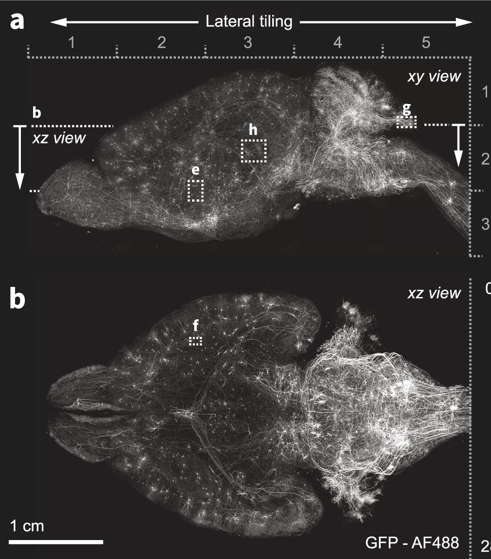
What it already does. ExA-SPIM images an entire expanded mouse brain in 15 tiles in as little as a day, without sectioning, at three-fold expansion, about 500 by 1,000 nanometers, or 375 by 750 nanometers at four-fold. With a sparse set of neurons labeled, each of them can be traced end to end across the brain, which is how complete single-neuron morphologies and projection atlases are built: a few neurons per brain, over many brains. That is real value, and it is the instrument we would start from.
Why it is not enough for a wiring diagram. The thinnest axons are 100 to 200 nanometers across and packed about 20 nanometers apart, so one 375 nanometer resolution element holds several of them. A tracer cannot follow one through the tangle or tell a synapse from a touch. Dense wiring, every neuron rather than a labeled few, has only been traced at about 20 to 30 nanometers: electron microscopy at 4 to 8, LICONN at an effective 10 by 10 by 25. The Allen instrument stops at 375 because it trades aperture for a 10 millimeter field and a 35 millimeter working distance, and resolution falls with aperture.
What sixteenfold adds. The same brain at LICONN's resolution holds every neuron, about 71 million, and every synapse, about 250 billion if MICrONS' count per cubic millimeter holds across the brain, with the proteins labeled, in one animal. A sparse brain answers one question about a few neurons. A dense brain answers the anatomical question for every neuron, once, and it is the map a model of the brain's wiring has to start from. Getting there means a light sheet at sixteenfold that holds LICONN's voxel over 64 times the volume, which has not been shown. That is the capital problem, and the one we are organized to solve. Neuron count: Herculano-Houzel et al., PNAS 2006; synapse density: MICrONS, 523 million per cubic millimeter.
Today's costs: Wellcome, 2023, with the per-cubic-millimeter figure our arithmetic on it. Our target is a goal. It has not been reached yet.